Br J Dermatol. 1999 Aug;141(2):224-30.
Source
INSERM U.346/CNRS ‘Human Skin and Immunity’, Hôpital E.Herriot, 69437 Lyon cedex 03, France. marek.haftek@lyon151.inserm.fr
Abstract
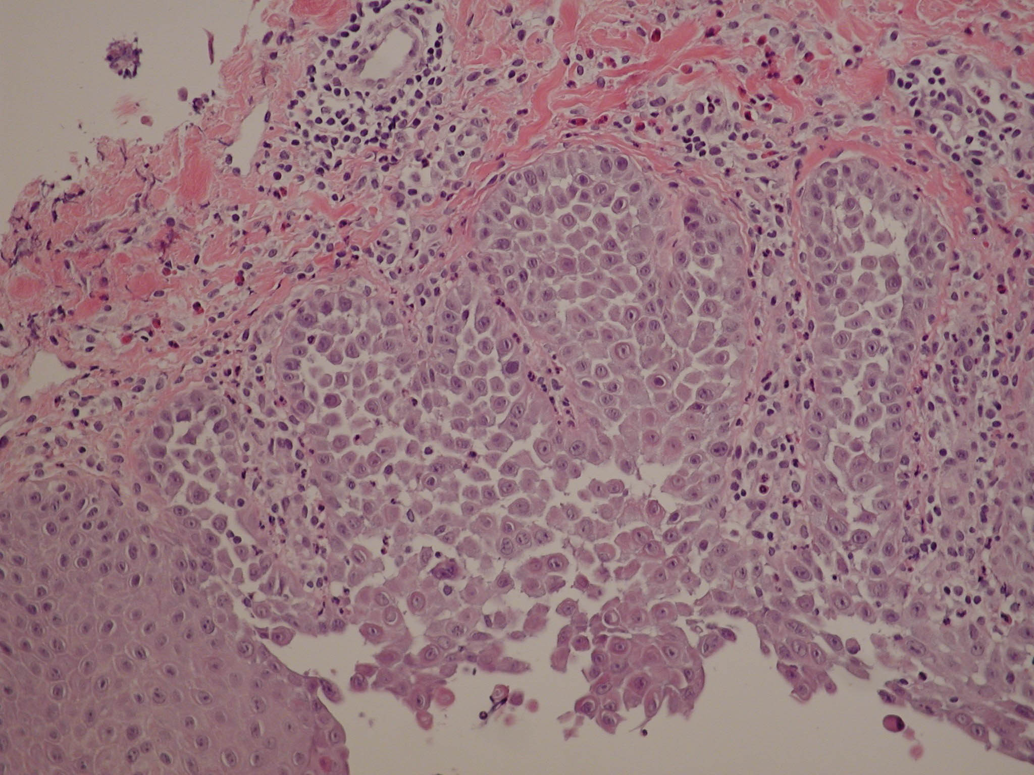
Hereditary skin disorders involving acantholysis, such as Hailey-Hailey disease and Darier’s disease, have been genetically linked to distinct chromosomal parts which do not code for known structural proteins. Such evidence suggests that the genomic abnormalities underlying these dermatoses may concern functional/regulatory mechanisms of keratinocyte cohesion. Epidermal communication junctions (gap junctions) are responsible for direct coupling of cells and, thus, co-ordinate the behaviour of keratinocytes within the tissue. Consequently, they remain one of the potential, and poorly studied, elements in the pathogenesis of hereditary acantholytic diseases. We have investigated the distribution and fate of gap junctions during non-immune acantholysis, using fine immunolocalization methods at the light and electron microscopic levels. Our results demonstrate normal expression of epidermal gap junction proteins, connexins 26 and 43, in non-lesional skin of Hailey-Hailey and Darier’s diseases. The gap junctions were not primarily dismantled during acantholysis, typical of both of the studied dermatoses, but underwent internalization and subsequent cytoplasmic dispersion in the portions of cells which were no longer attached to the rest of the tissue. In Darier’s disease, perifollicular acantholysis did not specifically concern epithelium of appendages coexpressing connexin 26 in addition to connexin 43, further indicating that the observed changes in gap junction localization were secondary to the loss of cell-cell contact. We demonstrated that the sequence of changes was identical in both diseases and that the previously described putative differences were apparently related to the degree of acantholysis present in the studied biopsies. The fate of the junctional structures and proteins, documented in the present study, is most probably a form of recycling process also used by normal keratinocytes during organogenesis and tissue differentiation.
