J Invest Dermatol. 2009 Jun;129(6):1379-87. doi: 10.1038/jid.2008.381. Epub 2008 Dec 4.
Source
Department of Anatomy and Cell Biology, Institute of Biomedicine, University of Oulu, Oulu, Finland. pekka.t.leinonen@oulu.fi
Abstract
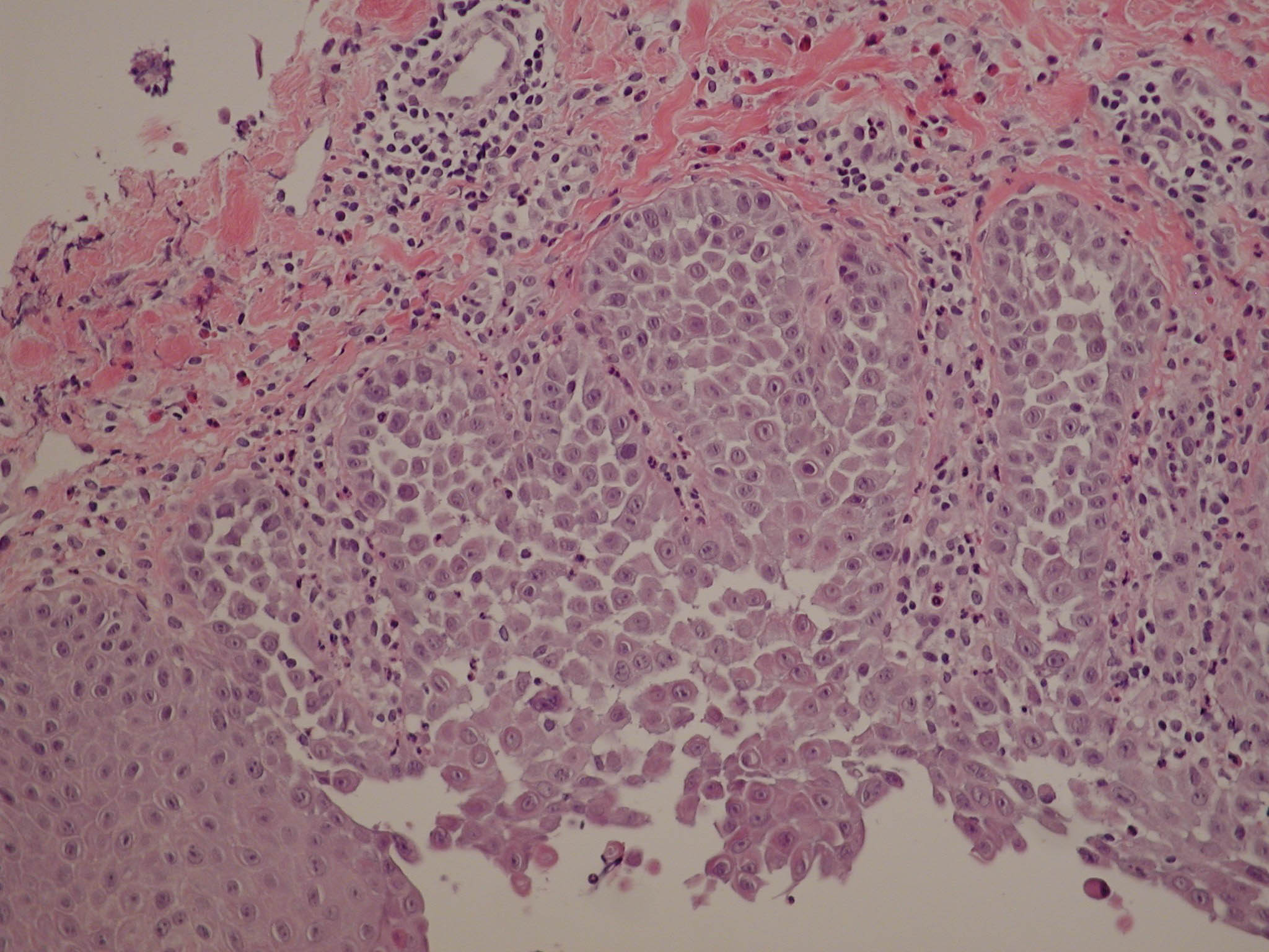
Electron probe microanalysis was used to analyze elemental content of human epidermis. The results revealed that the calcium content of the basal keratinocyte layer was higher than that of the lowest spinous cell layer in normal epidermis. This was surprising, as it is generally accepted that the calcium level increases with cellular differentiation from the proliferative basal layer to the stratum corneum. Hailey-Hailey disease (HHD) and Darier disease (DD) are caused by mutations in Ca(2+)-ATPases with the end result of desmosomal disruption and suprabasal acantholysis. The results demonstrated three major aberrations in HHD and DD lesions. First, in HHD and DD lesions the calcium content in the basal layer was lower than in the normal skin. Second, adenosine triphosphate (ATP) receptor P2Y2 was not localized to plasma membrane in acantholytic cells, whereas P2X7 appeared in the plasma membrane, potentially mediating apoptosis. Third, transition of keratin 14 to keratin 10 was abnormal as demonstrated by the presence of keratinocytes expressing both cytokeratins, which are usually exclusive in normal epidermis. Our results provide to our knowledge previously unreported elements for understanding how the disturbed calcium gradient is linked to the alterations in ATP receptors and keratin expression, leading to the clinical findings in HHD and DD.
